how to become more vascular
What are vascular studies?
(Carotid, Arm, and Leg Arterial and Venous Studies, Carotid Ultrasound, Venous Doppler Studies, Arterial Doppler Studies, Pulse Volume Recordings, PVRS)
Vascular studies are tests that check the blood flow in your arteries and veins. These tests are noninvasive. This means they don't use any needles.
Vascular studies use high-frequency sound waves (ultrasound) to measure the amount of blood flow in your blood vessels. A small handheld probe (transducer) is pressed against your skin. The sound waves move through your skin and other body tissues to the blood vessels. The sound waves echo off of the blood cells. These echoes are then sent to a computer and seen on a screen as images or video.
Vascular studies may use one of these special types of ultrasound technology:
-
Doppler ultrasound. This allows a healthcare provider to see blood flow through arteries and veins. The amount of blood pumped with each heartbeat is a sign of how large a vessel's opening is. A Doppler ultrasound can also find abnormal blood flow in a vessel, which may mean there is a blockage.
-
Color Doppler. This is an enhanced form of Doppler ultrasound. It uses different colors to show the direction of blood flow.

Why might I need a vascular study?
A vascular study may be done to:
-
Check signs and symptoms that may mean you have decreased blood flow in arteries or veins in your neck, legs, or arms
-
Assess procedures you have had done before to restore blood flow to an area
-
Assess a vascular dialysis device (such as an A-V fistula in the arm)
There may be other reasons for your doctor to recommend a vascular study.
Health problems that may cause decreased blood flow in arteries or veins include:
-
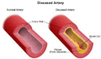
Atherosclerosis. A slow clogging of the arteries over many years by fatty materials (plaque) and other substances in the blood stream.
-
Aneurysm. An enlargement (dilation) of part of the heart muscle, or of the body's main artery (the aorta). This may cause tissue weakness at the aneurysm site.
-
Thrombus or embolus. A blood clot that forms in a blood vessel is a thrombus. An embolus is a small mass of material that moves through blood vessels to another part of the body, but gets stuck in a vessel.
-
Inflammatory conditions.Swelling (inflammation) in a blood vessel may occur because of an injury or an irritating medicine that gets into the vessel. It can also be caused by infection or an autoimmune disorder.
-
Varicose veins. Large, bulging veins in the leg. They occur when valves in the leg veins don't work well, allowing blood to collect in the lower leg.
Symptoms that may occur when blood flow decreases to your legs include:
-
Leg pain or weakness during exertion (claudication)
-
Swelling
-
Soreness, tenderness, redness, or warmth in the leg
-
Pale and cool skin, may even be a grayish or blue color
-
Numbness or tingling
-
Foot pain that occurs when sitting or lying down, and is relieved by standing (rest pain)
If your provider thinks you may have decreased blood flow in your arms, legs, or neck, then vascular studies may be done.
Types of vascular studies
There are different types of vascular studies. The tests that you have will depend on your symptoms and what your provider thinks your vascular problem may be. Types of vascular studies include:
Pulse volume recording (PVR) study. This is done to assess blood flow in your arms or legs. Blood pressure cuffs are inflated on your arm or leg, and the blood pressure there is measured using the Doppler probe.
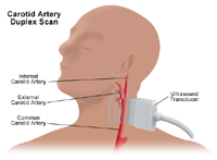
Carotid duplex scan. This type of Doppler exam checks the carotid arteries in your neck. It gives a 2D (2-dimensional) image of the arteries. This can show the structure of the arteries, the blocked area, and how well blood is flowing.
Carotid artery duplex scan. This type of vascular ultrasound can check for blockages or narrowing (stenosis) of the carotid arteries in your neck. It can also check the branches of the carotid artery.
Your healthcare provider may order other related tests or procedures, depending on your health problem.

What are the risks of a vascular study?
Vascular studies are safe and painless. They don't use radiation. And you will likely not feel any discomfort when the ultrasound probe is placed on your skin.
Some people may find it uncomfortable to have to lie still on the exam table for the procedure.
You may have other risks depending on your specific health problem. Be sure to discuss any concerns with your provider before the test.
Certain factors or conditions may interfere with a vascular study. These include:
-
Smoking for at least an hour before the test, as smoking causes blood vessels to tighten (constrict)
-
Severe obesity
-
Irregular heart rhythms (cardiac dysrhythmias or arrhythmias)
-
Heart disease
How do I get ready for a vascular study?
-
Your provider will explain the procedure to you. Ask any questions you have about the procedure.
-
Your healthcare provider may have other instructions for you based on your health problem.
-
Generally, you don't need to prepare for a vascular study.
-
Your doctor may give you specific instructions about smoking and having caffeine. You may be asked to not smoke for at least 2 hours before the test. This is because smoking makes blood vessels tighten (constrict). You may also be asked to not have caffeine in any form for about 2 hours before to the test.
What happens during a vascular study?
A vascular study may be done on an outpatient basis, which means you go home the same day. Or it may be done as part of a hospital stay. Procedures may vary depending on your condition and your provider's practices.
Generally, a vascular study follows this process:
-
You will be asked to remove any jewelry or other objects that may interfere with the procedure. You may wear your glasses, dentures, or hearing aid if you use any of these.
-
If you are asked to remove clothing, you will be given a gown to wear.
-
You will lie on an exam table or bed.
-
A clear gel will be placed on your skin at locations where the pulse is expected to be heard.
-
The Doppler probe will be pressed against your skin and moved around over the area of the artery or vein being studied.
-
When blood flow is detected, you will hear a "whoosh, whoosh" sound. The probe will be moved around to compare blood flow in different areas of the artery or vein.
-
For arterial studies of the legs, blood pressure cuffs will be used. They are put on 3 different places on your leg: your thigh, calf, and ankle. This is done to compare the blood pressure in these areas. The cuff around your thigh will be inflated first. The blood pressure is checked by putting the Doppler probe just below the cuff.
-
The cuff around your calf will be inflated, and the blood pressure checked.
-
The cuff around your ankle will be inflated, and the blood pressure checked.
-
Blood pressure is then taken in the arm that's on the same side as the leg that was just studied. This is used to find out how much the blood flow is blocked in your legs.
-
When the procedure is over, the gel will be removed from your skin.
What happens after a vascular study?
You may go back to your normal diet and activities unless your provider advises you differently.
Generally, there is no special type of care following a vascular study. Your healthcare provider may give you other instructions, depending on your situation.
how to become more vascular
Source: https://www.hopkinsmedicine.org/health/treatment-tests-and-therapies/vascular-studies
Posted by: danielswast1949.blogspot.com

0 Response to "how to become more vascular"
Post a Comment